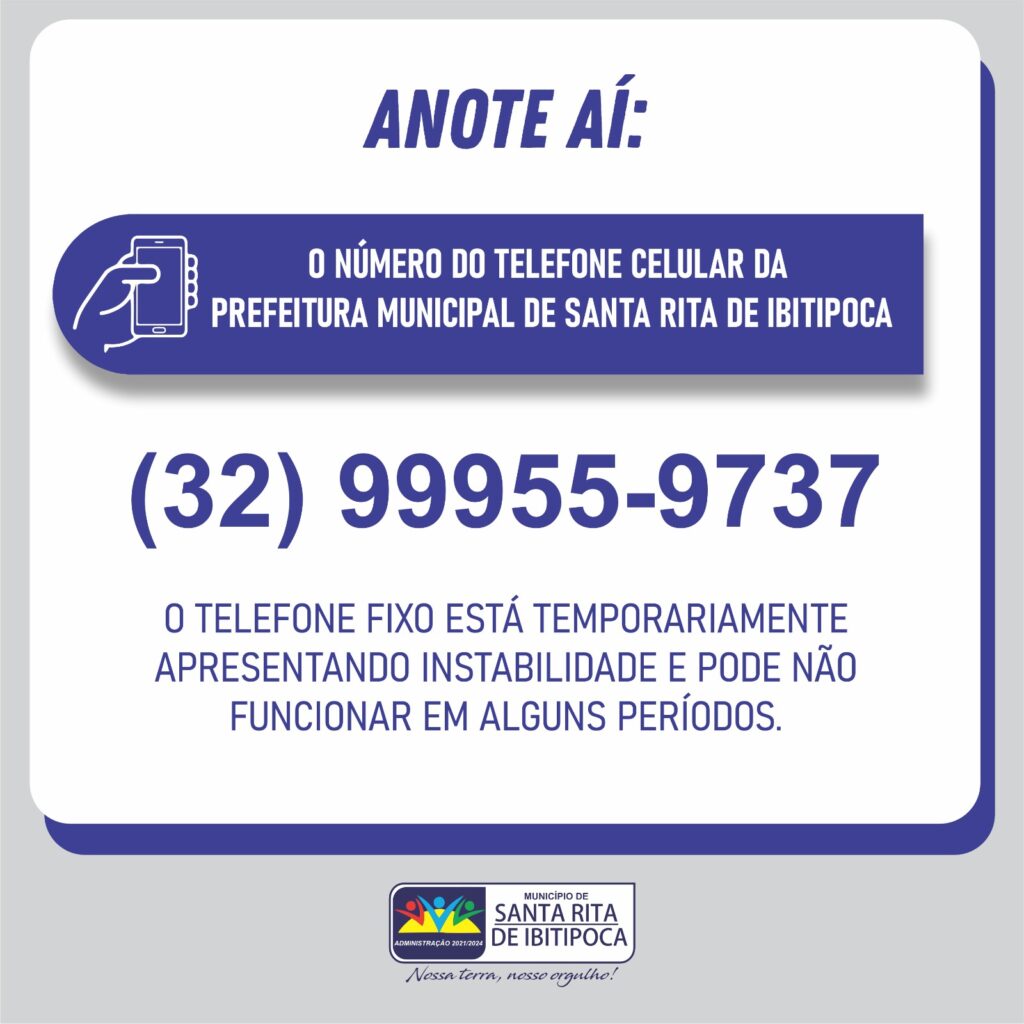
(32)99955-9737
Rua Francisco Novato, 02 – Centro – Santa Rita de Ibitipoca
gabinete@santaritadeibitipoca.mg.gov.br/
Search
Search
Início
Prefeitura
Gabinete
Estrutura Administrativa
Bens Culturais
Legislação
Código Tributário
Código de Posturas
LRF: RGF/RREO
PPA – LDO – LOA
Lei Orgânica
Decretos
Decretos Municipais 2020
Decretos Contábeis 2020
Decretos Municipais 2019
Decretos Contábeis 2019
Decretos Municipais 2018
Decretos Municipais 2017
Decretos Municipais 2016
Decretos Municipais 2015
Decretos Municipais 2014
Portarias
Portarias 2021
Portarias 2017
Portarias 2016
Portarias 2015
Portarias 2014
Leis Munipais
Leis Municipais 2023
Leis Municipais 2022
Leis Municipais 2021
Leis Municipais 2020
Leis Municipais 2019
Leis Municipais 2018
Leis Municipais 2017
Leis Municipais 2016
Folha de Pagamento
Diárias
Processos Licitatórios
Nova Lei – Licitações 2024
Editais de Licitação
Contratos 2020
Contratos 2018
Extratos de Contrato 2016
Extratos de Atas de Registro de Preço 2016
Extratos de Atas de Registro de Preço 2015
LAI
Convênios
Contato
Concurso
Webmail
Perguntas Frequentes
Convênios
Lei Paulo Gustavo
PNAB – ALDIR BLANC
Menu
Início
Prefeitura
Gabinete
Estrutura Administrativa
Bens Culturais
Legislação
Código Tributário
Código de Posturas
LRF: RGF/RREO
PPA – LDO – LOA
Lei Orgânica
Decretos
Decretos Municipais 2020
Decretos Contábeis 2020
Decretos Municipais 2019
Decretos Contábeis 2019
Decretos Municipais 2018
Decretos Municipais 2017
Decretos Municipais 2016
Decretos Municipais 2015
Decretos Municipais 2014
Portarias
Portarias 2021
Portarias 2017
Portarias 2016
Portarias 2015
Portarias 2014
Leis Munipais
Leis Municipais 2023
Leis Municipais 2022
Leis Municipais 2021
Leis Municipais 2020
Leis Municipais 2019
Leis Municipais 2018
Leis Municipais 2017
Leis Municipais 2016
Folha de Pagamento
Diárias
Processos Licitatórios
Nova Lei – Licitações 2024
Editais de Licitação
Contratos 2020
Contratos 2018
Extratos de Contrato 2016
Extratos de Atas de Registro de Preço 2016
Extratos de Atas de Registro de Preço 2015
LAI
Convênios
Contato
Concurso
Webmail
Perguntas Frequentes
Convênios
Lei Paulo Gustavo
PNAB – ALDIR BLANC
O que você procura?
Transparência
Destaques
Nova Lei de Licitações e Contratos, Lei nº 14.133/2021
Classificação do Edital para Cadastro de Professor Edital nº 001/2024
Edital para Recrutamento de Professor
NF Eletrônica
Acesso Rápido
Para acessar a todas as
Notícias
Clique aqui
Para acessar a todas as
Notícias
Clique aqui
© 2021 - Todos os Direitos Reservados
(32)98408-9581